Peer inside a mummified cat from ancient Egypt, courtesy of high-res 3D X-rays
Companies, agencies, institutions, etc
Scientific Reports
the Berlin Egyptian Museum
ReadingScientists
textNow
CT
Micro CT
Swansea University
the Egypt Centre
the Ars Orbital Transmission
CNMN Collection
WIRED Media Group
Condé Nast
People
Jennifer Ouellette
Leviticus
Ezekiel
Richard Johnston
Carolyn Graves-Brown
Ars
Groups
Egyptians
German
Eurasian
Physical locations
the Dead Sea
En Gedi
Places
No matching tags
Locations
CT
Egypt
Events
No matching tags
Summary
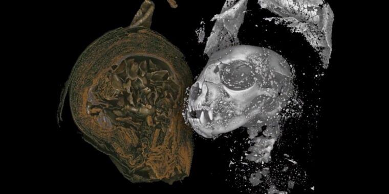
Now an inter-disciplinary team of scientists has managed to digitally "unwrap" three specimens—a mummified cat, bird, and snake—using a high-resolution 3D X-ray imaging technique, essentially enabling them to conduct a virtual postmortem, according to a new paper published in the journal Scientific Reports.Studying fragile ancient artifacts with cutting-edge imaging technology confers a powerful advantage on archaeological analysis. Previously, Micro CT had been used to image a mummified falcon, enabling researchers to determine its likely last meal and to image a mummified severed human hand."Using micro CT we can effectively carry out a postmortem on these animals, more than 2,000 years after they died in ancient Egypt," said co-author Richard Johnston of Swansea University. Our work shows how the hi-tech tools of today can shed new light on the distant past."Swansea University maintains a collection of mummified specimens, and the team selected three animal remains that varied in both size and shape: a cat, a bird, and a snake. The mummified remains of the snake were not definitely identified as a snake until a 2009 radiograph, courtesy of a local veterinary clinic, revealed it to be coiled up inside.Using micro CT, the team was able to image the remains in extraordinary detail, including small bones and teeth, desiccated soft tissues, and mummification materials.
As said here by Jennifer Ouellette